Advanced Diagnostics with Computed Tomography (CT) – Nasal Tumor Case Study
Case Studies

Mosh, a 5-year-old male domestic short hair presented to Pet Emergency & Referral Center (PERC) with nasal congestion, left-sided nasal discharge, and left-sided epistaxis. This had been going on at home for 2-3 weeks.
Previously Mosh underwent diagnostic lab work at his primary veterinarian which revealed mild leukocytosis, with mature neutrophilia. The skull and thoracic radiographs revealed opacification of the left nasal cavity without visible osteolysis or displacement of the nasal septum. Mosh was given antibiotics, Cerenia intranasal drops to reduce nasal discharge, and l-lysine amino acids.
Unfortunately, symptoms continued to progress and it became difficult for Mosh to breathe from the severe congestion. At this point, his veterinarian referred him to PERC for further testing.
Internal Medicine Specialist, Beth Lechner, DVM, DACVIM, performed an urgent CT scan and Rhinoscopy.
CT Report Findings:
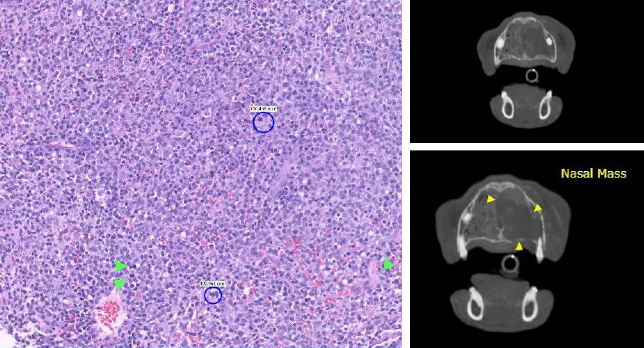
A left nasal soft tissue mass with polyostotic aggressive osteolytic lesions of the osseous lining of the left nasal cavity
Secondary complete upper airway obstruction
Secondary left-sided obstructive sinusitis
Lymphadenopathy mandibular lymph nodes
Absent triadan 101 and 201.
The left nasal mass was consistent with primary nasal neoplasia and secondary aggressive osteolytic lesions of the associated osseous structures. Differentials included lymphosarcoma or less likely adenocarcinoma, squamous cell carcinoma, or transitional cell carcinoma.
Dr. Lechner performed a rhinoscopy biopsy which confirmed the Adam tumor stage was T3. A fine needle aspirate sample of the tributary lymph nodes was performed to screen for metastatic disease.
Rhinoscopy Report Findings:
Retroflex nasopharyngoscopy: A large and pale mass was noted at the choana, obstructing much of the choanal opening. Multiple raised soft tissue nodules found along the dorsal soft palate, suspected to represent prominent lymphoid tissue
Antegrade rhinoscopy: Impaired visualization of the left nare due to increased discharge and mass effect. Right nare was mildly erythematous but had smooth mucosa
Biopsies were collected from right and left nares
Histopathology Report:
Left nasal turbinates: Favored lymphoma, intermediate to large cell Mitotic count: 18 mitotic figures per 10 high-powered fields Margins: Incisional sections
Vascular invasion: None were observed in right or left nasal turbinates: Moderate lymphoplasmacytic and neutrophilic rhinitis with rare suspected surface bacteria
Mosh was referred for consultation with oncology to discuss treatment options for the treatment of nasal lymphoma (LSA) with radiation therapy and chemotherapy.
